How to draw a heart given to people. Anatomy and physiology of the heart: structure, functions, hemodynamics, cardiac cycle, morphology
Want to know How to draw human heart pencil step by step, take a few simple steps.
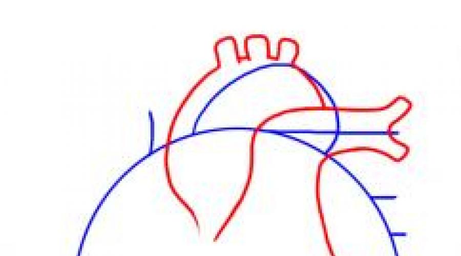
Step 1. Okay, let's start this lesson on the human heart, should we? First sketch out some guidelines and shapes so that we have a nice workable wireframe to use. Start with a circle for the heart, and then draw the lower part of the heart that contains the heart muscle. The three horizontal lines you see drawn will be plugs on the pulmonary artery, pulmonary veins, and the left atrium. The lump you see drawn will be for the aorta.

Step 2. Okay, sketch out the actual shapes of the aorta as well as the tubes that come from that part of the heart. You will then sketch out the shape for the pulmonary artery as you see here.

Step 3. Now, sketch out the shape of the vena cava, which is in the vase looking at the tube, which is all by itself. Next draw four tubes. The upper tube enters the pulmonary artery, and the last three are the pulmonary veins, which are on the left side. Next, sketch out the outer shape of the heart on the left side, which is also part of the heart muscle, and then draw in the veins that lie on the surface of the heart. Finally, you will draw a tube for the inferior vena cava, which is just below the lower left side of the heart.

Step 4. This is your last drawing step and all you need to do is draw out the remaining actual human heart shape and then draw in the superficial veins. Finally, sketch out the tubes for the pulmonary vein and left atrium. Erase all guidelines that are visible to clear your drawing of a human heart.
The heart means a lot to a person. A real heart is the basis of our body, and valentines or simple drawn hearts help to express our feelings. This is a manifestation of warmth, love and tender feelings for a person. Below we give a few simple tips how to draw a heart. There are several options for drawing, you can use them or come up with your own.
Simplified version
Before you draw a heart with a pencil (or rather start), prepare all the tools (paper, eraser, pencils). Place a piece of paper in front of you. First you need to think over the details if you want to add something to the heart. Make sure all parts of the drawing fit on the sheet. Better schematically (in squares, circles) to draw all the main elements. Now we take a pencil and proceed. There are three options for how to draw a valentine heart

The first way
Put a dot in the center of the sheet, it will be the base of the heart. Draw a semicircular line, directing it first up to the right and then down. The end point of the arc must be below the base point. You should end up with something that looks like a question mark. Repeat the steps on the left half of the sheet. The lines should converge at one point.
Second way
Draw inverted isosceles triangle(the base should be at the top). Draw a bisector from the bottom vertex. Then "write" in each of the resulting triangles half a heart. Use the eraser to remove unnecessary lines.
The third way
Draw two intersecting circles (you can use stencils) and draw a heart based on them. If you get it asymmetrical, then fold a sheet of paper in half and draw one half at the fold line, then cut it out. Now you know how to draw a beautiful heart in its simplified form. When you have the base ready, you can use your imagination: pierce the heart with arrows, thorns, draw around a rose or wings. You can color it in or circle it with a marker, leaving it in black and white. Do not overload the drawing with a lot of unnecessary details.
How to draw a human heart
You will also need tools, prepare yourself a space. Better to use a vertically oriented sheet. In this case, you need to study well the anatomy of the human heart. Can be copied from a textbook or medical reference book.

Brief description of the process:
You need to draw an oval that tapers downward. It should be slightly tilted. Then draw the right atrium. An important part of the heart is the aorta, don't forget about it. This is a large "tube" that will be located in the upper part of the figure, three more vessels emerge from it. Add veins, don't forget the left atrium. Also circle the picture and color if desired. Don't forget to erase any extra lines.
Conclusion
Now you know several ways to draw a heart. If you're having trouble drawing, don't give up. When everything starts to work out for you, you can please your loved one with a beautiful handmade valentine.
When there is an eternal spring in the soul, the elated mood cannot be restrained in any way: it just bursts out of the chest in order to splash out with cute creativity. How to draw and better - both together? Take a simple pencil clear sheet papers - now you will find out everything.
Lesson # 1: How to draw a heart with a pencil
We will draw a heart from roses. Draw a regular circle and divide it in half with a line. Exactly on horizontal line depict an uneven oval, similar to a deflated ball. Attach a pair of curves to it at the top and bottom, like the red lines shown in the illustration for an example.

In the lesson explaining how to draw a heart, first of all, pay attention to the red lines of the pattern - these are new fragments that need to be repeated on your version of the original heart.

Draw a kind of snail in the very center of the future masterpiece. First, simply divide the uneven oval almost in half with a convex curved line. Add a few strokes: in the form of the letter "P" and from its top a regular line, bounded by the same oval. Don't forget to add one more stroke, very small, in the top petal. This inverted "comma" will add volume to the drawing.

Quite a simple step creative process titled "How to draw a heart": draw two symmetrical petals, the upper part of which, as it were, repeats the invisible lines of the heart.

All that's left is to add three petals at the bottom of the heart. If you haven't skipped your math class at school, you can draw curly braces. This acquired skill will help you easily cope with the task: a pair of curves to the left and right and another, final one, with an “arrow” down the center. By the way, looking closely at the sample, you will see that the last petal you draw will be boring without a small detail - a convex stroke that adds volume.

Erase all auxiliary, erroneous and unnecessary lines. Completed the lesson "How to Draw a Heart"!
Lesson # 2: Heart surrounded by roses
Let's complicate the task: let's draw in a scarlet round dance:

Draw an arbitrary outline of the heart, for example, such as here:

Make the first sketches of three buds at once, spreading them evenly. Start with the curls, from them draw the side lines in the pattern:

Each flower has its own individual shape, which appears thanks to simple curved lines. Take a closer look and repeat them in your drawing, there is nothing complicated about it:

Let's complete the drawing of the roses by adding three to four graceful contours to each bud.
The heart is a muscular organ in humans and animals that pumps blood through the blood vessels.
Heart functions - why do we need a heart?
Our blood provides the entire body with oxygen and nutrients. In addition, it also has a cleansing function, helping to remove metabolic waste.
The heart's function is to pump blood through the blood vessels.
How much blood does the human heart pump?
The human heart pumps from 7,000 to 10,000 liters of blood in one day. This amounts to approximately 3 million liters per year. It turns out up to 200 million liters in a lifetime!
The amount of blood pumped over a minute depends on the current physical and emotional load - the greater the load, the more blood the body needs. So the heart can pass through itself from 5 to 30 liters in one minute.
The circulatory system consists of about 65 thousand vessels, their total length is about 100 thousand kilometers! Yes, we have not sealed ourselves.
Circulatory system
The human cardiovascular system is formed by two circles of blood circulation. With each heartbeat, blood moves in both circles at once.
Small circle of blood circulation
- Deoxygenated blood from the superior and inferior vena cava enters the right atrium and further into the right ventricle.
- From the right ventricle, blood is pushed into the pulmonary trunk. The pulmonary arteries conduct blood directly to the lungs (up to the pulmonary capillaries), where it receives oxygen and gives off carbon dioxide.
- Having received enough oxygen, blood returns to the left atrium of the heart through the pulmonary veins.
A large circle of blood circulation
- From the left atrium, blood moves into the left ventricle, from where it is further pumped out through the aorta into the systemic circulation.
- Having passed a difficult path, blood through the vena cava again arrives at the right atrium of the heart.
Normally, the amount of blood expelled from the ventricles of the heart is the same with each contraction. So, an equal volume of blood flows into the large and small circles of blood circulation at the same time.
What is the difference between veins and arteries?
- The veins are designed to transport blood to the heart, while the arteries are designed to deliver blood in the opposite direction.
- The blood pressure in the veins is lower than in the arteries. Accordingly, the walls of the arteries are distinguished by greater extensibility and density.
- Arteries saturate "fresh" tissue, and veins take "waste" blood.
- In case of vascular damage, arterial or venous bleeding can be distinguished by its intensity and blood color. Arterial - strong, pulsating, beating like a "fountain", the color of the blood is bright. Venous - bleeding of constant intensity (continuous flow), the color of the blood is dark.
The weight of a human heart is only about 300 grams (on average 250g for women and 330g for men). Despite its relatively low weight, it is undoubtedly the main muscle in the human body and the basis of its life. The size of the heart is indeed approximately equal to the fist of a person. Athletes can have a heart one and a half times larger than that of an ordinary person.
Anatomical structure
The heart is in the middle chest at the level of 5-8 vertebrae.
Fine, Bottom part the heart is located mostly in the left half of the chest. There is a variant of congenital pathology in which all organs are mirrored. It is called transposition internal organs... The lung, next to which the heart is located (normally - the left), has a smaller size relative to the other half.
The posterior surface of the heart is located near the spinal column, and the anterior surface is reliably protected by the sternum and ribs.
The human heart consists of four independent cavities (chambers) divided by partitions:
- the upper two - the left and right atria;
- and two lower - left and right ventricles.
The right side of the heart includes the right atrium and ventricle. The left half of the heart is represented by the left ventricle and atrium, respectively.
The inferior and superior vena cava enter the right atrium, and the pulmonary veins enter the left. From right ventricle the pulmonary arteries (also called the pulmonary trunk) come out. From left ventricle the ascending aorta rises.
The heart has protection from overstretching and other organs, which is called the pericardium or pericardial sac (a kind of shell, which encloses the organ). Has two layers: an outer dense, strong connective tissue called fibrous membrane of the pericardium and internal ( serous pericardium).
Thus, the heart itself consists of three layers: epicardium, myocardium, endocardium. It is the contraction of the myocardium that pumps blood through the vessels of the body.
The walls of the left ventricle are about three times larger than the walls of the right! Explained given fact the fact that the function of the left ventricle is to push blood into the systemic circulation, where the opposition and pressure are much higher than in the small.
Heart valve device
Special heart valves allow the blood flow to be constantly maintained in the correct (unidirectional) direction. The valves open and close in turn, letting in blood, then blocking its path. Interestingly, all four valves are located along the same plane.
Between the right atrium and the right ventricle is located tricuspid (tricuspid) valve. It contains three special leaflet plates that, during the contraction of the right ventricle, are able to protect against the return flow (regurgitation) of blood into the atrium.
Works in a similar way mitral valve, only it is located on the left side of the heart and is bicuspid in structure.
Aortic valve prevents the backflow of blood from the aorta to the left ventricle. Interestingly, when the left ventricle contracts, the aortic valve opens as a result of blood pressure on it, so it moves into the aorta. Then, during diastole (the period of relaxation of the heart), the reverse flow of blood from the artery helps to close the valves.
Normally, the aortic valve has three cusps. The most common congenital heart anomaly is bicuspid aortic valve. This pathology occurs in 2% of the human population.
Pulmonary (pulmonary) valve at the moment of contraction of the right ventricle, it allows blood to flow into the pulmonary trunk, and during diastole it does not allow it to flow in the opposite direction. Also consists of three leaves.
Heart vessels and coronary circulation
 The human heart needs nutrition and oxygen, just like any other organ. The vessels supplying (feeding) the heart with blood are called coronary or coronary... These vessels branch off from the base of the aorta.
The coronary arteries supply the heart with blood, and the coronary veins carry deoxygenated blood. Those arteries that are on the surface of the heart are called epicardial. Subendocardial arteries are called coronary arteries hidden deep in the myocardium.
Most of the outflow of blood from the myocardium occurs through three cardiac veins: large, medium and small. Forming the coronary sinus, they flow into the right atrium. The anterior and lesser veins of the heart deliver blood directly to the right atrium.
Coronary arteries are classified into two types - right and left. The latter consists of the anterior interventricular and circumflex arteries. The great heart vein branches into the posterior, middle and small veins of the heart.
Even absolutely healthy people have their own unique features of coronary circulation. In reality, the vessels may look and be located differently than shown in the picture.
How does the heart develop (form)?
Impulse path
This system ensures the automatism of the heart - the excitation of impulses born in cardiomyocytes without external stimulus. In a healthy heart main source impulses - sinoatrial (sinus) node. He is the leader and blocks impulses from all other pacemakers. But if any disease occurs that leads to sick sinus syndrome, then other parts of the heart take over its function. So the atrioventricular node (automatic center of the second order) and the bundle of His (AC of the third order) are able to activate when the sinus node is weak. There are cases when secondary nodes enhance their own automatism during normal operation of the sinus node.
Sinus node located in the upper posterior wall of the right atrium in the immediate vicinity of the mouth of the superior vena cava. This node initiates pulses at a rate of approximately 80-100 times per minute.
Atrioventricular node (AV) located in the lower part of the right atrium in the atrioventricular septum. This septum prevents the propagation of the impulse directly into the ventricles, bypassing the AV node. If the sinus node is weakened, then the atrioventricular node will take over its function and begin to transmit impulses to the heart muscle with a frequency of 40-60 beats per minute.
Further, the atrioventricular node goes into bundle of His(the atrioventricular bundle is subdivided into two legs). The right leg rushes to the right ventricle. The left leg is divided into two more halves.
The situation with the left bundle branch is not fully understood. It is believed that the left leg with the fibers of the anterior branch rushes to the anterior and lateral walls of the left ventricle, and the posterior branch supplies fibers to the posterior wall of the left ventricle and the lower parts of the lateral wall.
In case of weakness of the sinus node and atrioventricular blockade, the His bundle is able to create impulses at a speed of 30-40 per minute.
The conducting system deepens and further branching into smaller branches, eventually passing into Purkinje fibers, which penetrate the entire myocardium and serve as a transmission mechanism for the contraction of the ventricular muscles. Purkinje fibers are capable of initiating pulses with a frequency of 15-20 per minute.
Exceptionally trained athletes can have a normal resting heart rate down to the lowest on record - as little as 28 beats per minute! However, for the average person, even if they lead a very active lifestyle, a heart rate below 50 beats per minute can be a sign of bradycardia. If you have such a low heart rate, then you should be examined by a cardiologist.
Heartbeat
A newborn's heart rate can be around 120 beats per minute. Growing up pulse an ordinary person stabilizes in the range from 60 to 100 beats per minute. Well-trained athletes ( it comes about people with well-trained cardiovascular and respiratory systems) have a pulse of 40 to 100 beats per minute.
The rhythm of the heart is controlled nervous system- sympathetic enhances contractions, and parasympathetic weakens.
Cardiac activity, to a certain extent, depends on the content of calcium and potassium ions in the blood. Other biologically active substances also contribute to the regulation of the heart rhythm. Our heart can begin to beat faster under the influence of endorphins and hormones released when listening to your favorite music or kissing.
In addition, the endocrine system is able to significantly influence the heart rate - both the frequency of contractions and their strength. For example, the release of the adrenal glands by the well-known adrenaline causes an increase in heart rate. The opposite hormone is acetylcholine.
Heart tones
One of the most simple methods Diagnosing heart disease is listening to the chest with a stethophonendoscope (auscultation).
In a healthy heart, with standard auscultation, only two heart sounds are heard - they are called S1 and S2:
- S1 - the sound heard when the atrioventricular (mitral and tricuspid) valves are closed during systole (contraction) of the ventricles.
- S2 - the sound heard when the semilunar (aortic and pulmonary) valves close during diastole (relaxation) of the ventricles.
Each sound has two components, but for the human ear they merge into one due to the very small time interval between them. If, under normal conditions of auscultation, additional tones are heard, then this may indicate some kind of disease of the cardiovascular system.
Sometimes, additional abnormal sounds called heart murmurs can be heard in the heart. As a rule, the presence of murmurs indicates some kind of heart pathology. For example, a murmur can cause blood to return in the opposite direction (regurgitation) due to malfunctioning or damage to a valve. However, noise is not always a symptom of the disease. To clarify the reasons for the appearance of additional sounds in the heart, it is worth doing echocardiography (ultrasound of the heart).
Heart disease
Not surprisingly, the number of cardiovascular diseases is on the increase in the world. The heart is a complex organ that actually rests (if you can call it rest) only in the intervals between heartbeats. Any complex and constantly working mechanism itself requires as much respectful attitude and constant prevention.
Just imagine what a terrible burden falls on the heart, given our lifestyle and poor quality plentiful nutrition. Interestingly, mortality from cardiovascular diseases is also quite high in countries with high level income.
The huge amounts of food consumed by the population of wealthy countries and the endless pursuit of money, as well as the stress associated with this, destroy our hearts. Another reason for the spread of cardiovascular diseases is physical inactivity - catastrophically low physical activity, destroying the entire body. Or, on the contrary, an illiterate passion for heavy physical exercise, often occurring against a background, the presence of which people do not even suspect and manage to die right during the "health-improving" activities.
Lifestyle and heart health
The main factors that increase the risk of developing cardiovascular disease are:
- Obesity.
- High blood pressure.
- Increased blood cholesterol levels.
- Physical inactivity or excessive physical activity.
- Abundant poor quality food.
- Suppressed emotional state and stress.
Take a read of this great article a turning point in your life - give up bad habits and change your lifestyle.
Heart, cor, represents a hollow muscular organ that receives blood from the venous trunks pouring into it and drives blood into the arterial system. The heart cavity is divided into 4 chambers: 2 atria and 2 ventricles. The left atrium and the left ventricle together constitute the left, or arterial, heart by the property of the blood in it; the right atrium and the right ventricle make up the right, or venous, heart. The contraction of the walls of the heart chambers is called systole, relaxing them - diastole.
The heart has the shape of a somewhat flattened cone. It distinguishes top, apex, base, basis, anterosuperior and lower surfaces and two edges - right and left, dividing these surfaces.
Rounded apex of the heart, apex cordis, facing down, forward and to the left, reaching the fifth intercostal space at a distance of 8 - 9 cm to the left of the midline; the apex of the heart is formed entirely by the left ventricle. Basis, basis cordis, facing up, back and right. It is formed by the atria, and in front - by the aorta and pulmonary trunk. In the upper right corner of the quadrangle formed by the atria, there is a place - the entry of the superior vena cava, in the lower - the inferior vena cava; now to the left are the places of entry of two right pulmonary veins, on the left edge of the base - two left pulmonary veins. Anterior, or sternocostal, surface of the heart, facies sternocostalis... facing anteriorly, upward and to the left and lies behind the body of the sternum and cartilage of the ribs from III to VI. The coronary sulcus, sulcus coronarius, which runs transversely to the longitudinal axis of the heart and separates the atria from the ventricles, the heart is divided into an upper section, formed by the atria, and into a larger lower section, formed by the ventricles. Walking on facies sternocostalis anterior longitudinal groove, sulcus interventricularis anterior... runs along the border between the ventricles, and most the anterior surface forms the right ventricle, the smaller - the left one.
The lower, or diaphragmatic, surface, facies diaphragmatica, adjacent to the diaphragm, to its tendon center. On it passes posterior longitudinal groove, sulcus interventricularis posterior... which separates the surface of the left ventricle (large) from the surface of the right (smaller). The anterior and posterior interventricular grooves of the heart with their lower ends merge with each other and form on the right edge of the heart, immediately to the right of the apex of the heart, heart tenderloin, incisura apicis cordis. The edges of the heart, right and left, of unequal configuration: the right is more acute; the left edge is rounded, more blunt due to the greater thickness of the wall of the left ventricle.

It is believed that the heart is equal in size fist of the respective individual... Its average dimensions are: length 12-13 cm, maximum diameter 9-10.5 cm, anteroposterior size 6-7 cm.The weight of a man's heart is on average 300 g (1/215 body weight), women - 220 g (1/250 body weight).
Anatomy of the heart (illustrations, 3D images, section photos)
Images and anatomical references
Human heart, anatomy and physiology

Human heart is a muscle pump that has been affecting the minds of people for hundreds of years. In 2725. BC e. in Egypt, Imhotep concluded that the pulse was associated with cardiac function. In 400g. BC e. Hippocrates wrote about the heart as a strong muscle.
In 1628. William Harvey has published an explanation of the circulatory process. Between 1857 and 1882, Marey and Dudgeon, independently of each other, created an apparatus for measuring blood pressure when hypertensive disease was detected in a person.
V last years molecular biology helped discover even more complex functions of this engineering masterpiece - human heart... which confirms the words of the psalmist that you and I are "wonderfully made" (Psalm 139: 14).
The term "cardiovascular" describes the heart and blood vessels of the body. The blood vessels are also sometimes called the vascular tree, or bed. In this article, we will look at the structure and functions human hearts .
The heart is a hollow muscular organ that sits in the center of the chest, but most of it is to the left of the midline.
Human heart consists of two upper chambers called the atria and two lower chambers called the ventricles. Structurally and functionally, the heart is divided into right and left parts; the right side pumps blood to the lungs, the left side throughout the body.
The upper chamber, or atrium, collects blood and pumps it into the ventricle, which then expels it from human heart into large vessels. There are inlet and outlet valves in each ventricle to provide blood flow in one direction.
Left ventricle.
Blood enters the left ventricle from the left atrium through the mitral valve, which is made up of two large cusps that open when the ventricle is relaxed (diastole).
When the filling of the ventricle is over and it contracts, the force of contraction "presses" the blood to the lower part of the mitral valve leaflets, as a result of which the valve closes. Thanks to this mechanism, blood flows in one direction - from the ventricle to the aorta.
The outlet valve of the left ventricle is called the aortic valve. It has three leaves, or cusps, that open when the ventricle contracts, allowing blood to enter the systemic circulation.
As the ventricle relaxes and the pressure drops below the pressure in the aorta, blood begins to flow back (from the aorta to the ventricle).
This reverse flow of blood causes the aortic valve leaflets to fill from above and thus approach each other (touch each other) and close. The valve closes and there is no reverse flow of blood into the left ventricle.
Right ventricle.
The inlet valve is a tricuspid valve, which, by definition, has three leaves. It provides unilateral blood flow from the right atrium to the right ventricle.
The blood is then released into the pulmonary artery through the pulmonary valve (consisting of three cusps) and flows to the lungs. The tricuspid and pulmonary valves close and open in the same way as the mitral and aortic valves, respectively.
The leaflets of the mitral and tricuspid valves are tied to the walls of the ventricles by "cords" of tissue and muscle called tendon filaments (chords) and papillary (papillary) muscles.
These structures keep the valves from opening in reverse side, which would cause the blood to flow in the opposite direction. If these valves, threads or muscles are damaged due to painful processes, then the valves do not close completely and may "leak" (valve failure).
There are also diseases that cause the valves to narrow, which in turn causes the blood flow through the valves to decrease.
As a result human heart the increased resistance is constantly overcome and it increases in size. However, over time, it depletes its reserve of strength and can no longer pump blood so effectively, which affects the health of the whole body.
The valves can also be affected by both processes simultaneously (constriction and "leakage"), as a result of which cardiac function is weakened and blood circulation is impaired.
Cardiac function.
The cardiac function consists in pumping blood through the large circle of blood circulation (the whole body) and through the small (pulmonary). The right side of the heart pumps blood to the lungs, where carbon dioxide is extracted from it and it is saturated with oxygen.
The left side of the heart pumps blood to the rest of the organs; thus oxygen and nutrients are delivered to them. Waste also enters the bloodstream, but this time venous, so that later they are removed from the body by such organs as the lungs, kidneys and liver.
The contraction and relaxation of the heart is a cardiac cycle that can be felt by feeling the pulsation of blood flowing through the arteries. This can be done by pressing the arteries against the bone, for example, in the wrist, lower leg, neck.
Arterial pulsation is created by a build-up of a pressure wave that flows through human arteries from the heart and causes a pulsating expansion of the arterial walls. If we count this pulsation for 60 s, then we get the pulse rate. In a healthy adult, it is approximately 72 beats per minute (the normal range is 65 to 90).
Each cardiac cycle consists of two phases: diastole and systole.
Diastole (or relaxation of the heart muscle) During this phase, the heart muscle relaxes in order to take in some volume flowing into the lumen heart, human blood. The atria then contract to move blood to the ventricles.
The next phase is called systole, or ventricular contraction, during which blood is pumped out of the heart. The atria begin to relax in order to receive more blood to repeat the cycle.
You can not only feel the pulse, but also follow the heart cycle if you listen to heart sounds through the chest wall using a stethoscope. These sounds are described as “lab-dub”, where the first “lab” sound indicates the closure of the mitral and tricuspid valves, and the second “dub” sound indicates the aortic and pulmonary valves.
Additional sounds usually indicate some kind of heart valve and / or muscle function abnormality. The most common sounds that indicate valve dysfunction are called murmurs.
These sounds are produced when turbulent blood flow occurs due to structural changes in the valve apparatus. Normally, the blood flow is smooth, linear and non-turbulent (non-vortex).
Electrical activity of the heart in humans.
In order for the heart to beat in an orderly fashion, it is equipped with nerve pacemakers (an accumulation of nerve cells in the atria) and a special conduction system that delivers nerve impulses to the heart muscle.
Different parts of the conduction system and even parts of the heart itself are capable of beating at different rates. The conduction system provides consistent, coordinated activation from the atria to the ventricles.
This electrical system ensures that the impulses reach all parts of the heart muscle. The electrical axis of the heart is determined using electrocardiogram (ECG) data.